


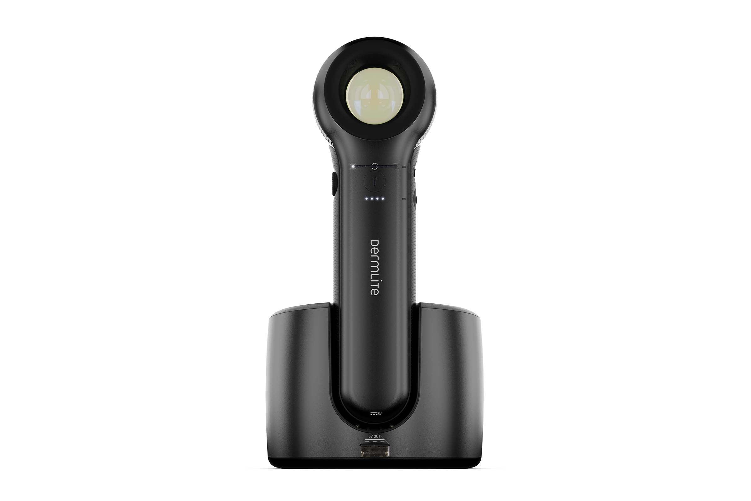
DermLite DL 5
- Regular price
-
€2.140,00 - Regular price
-
- Sale price
-
€2.140,00
Shipping only EU-wide
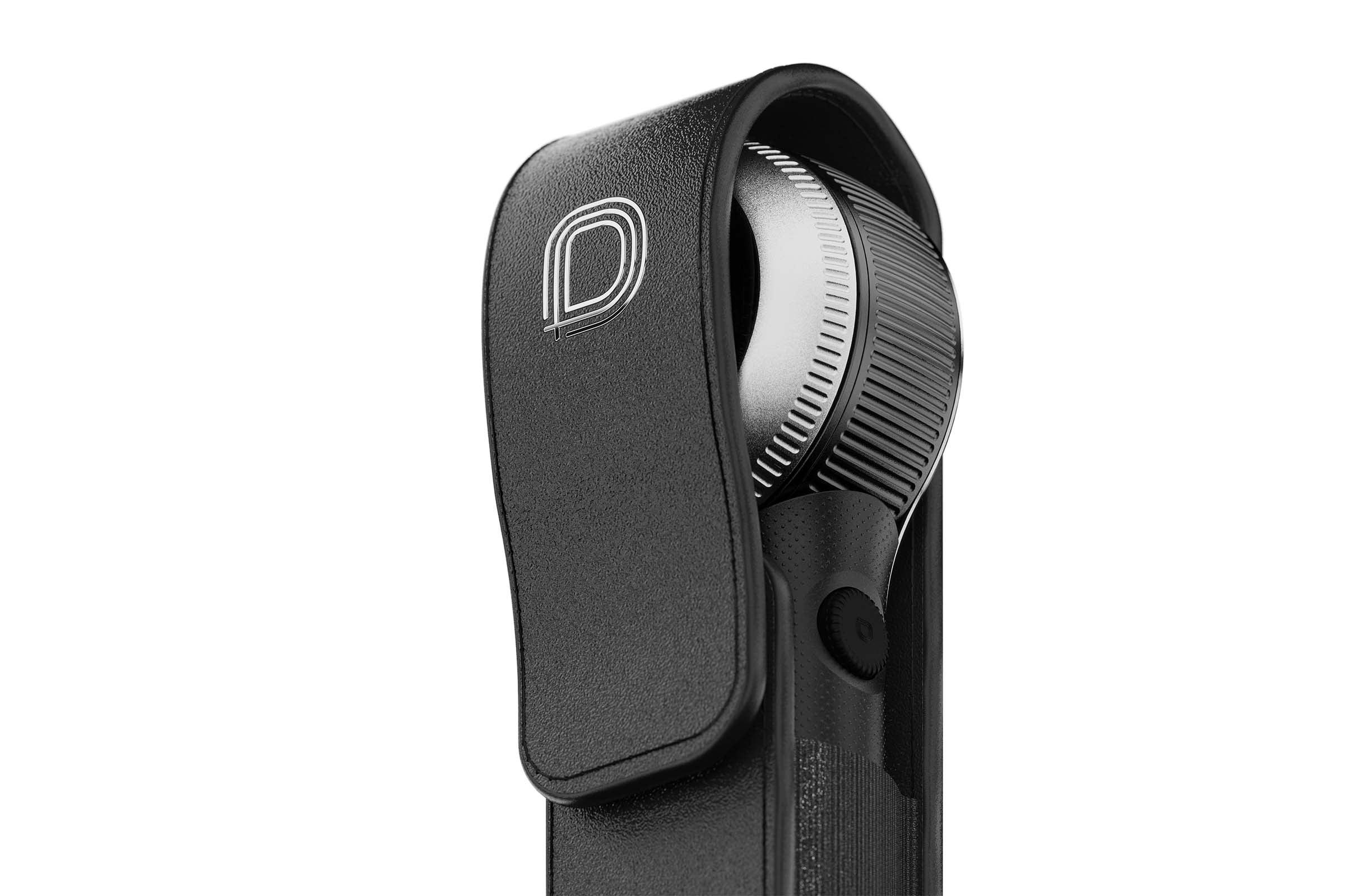
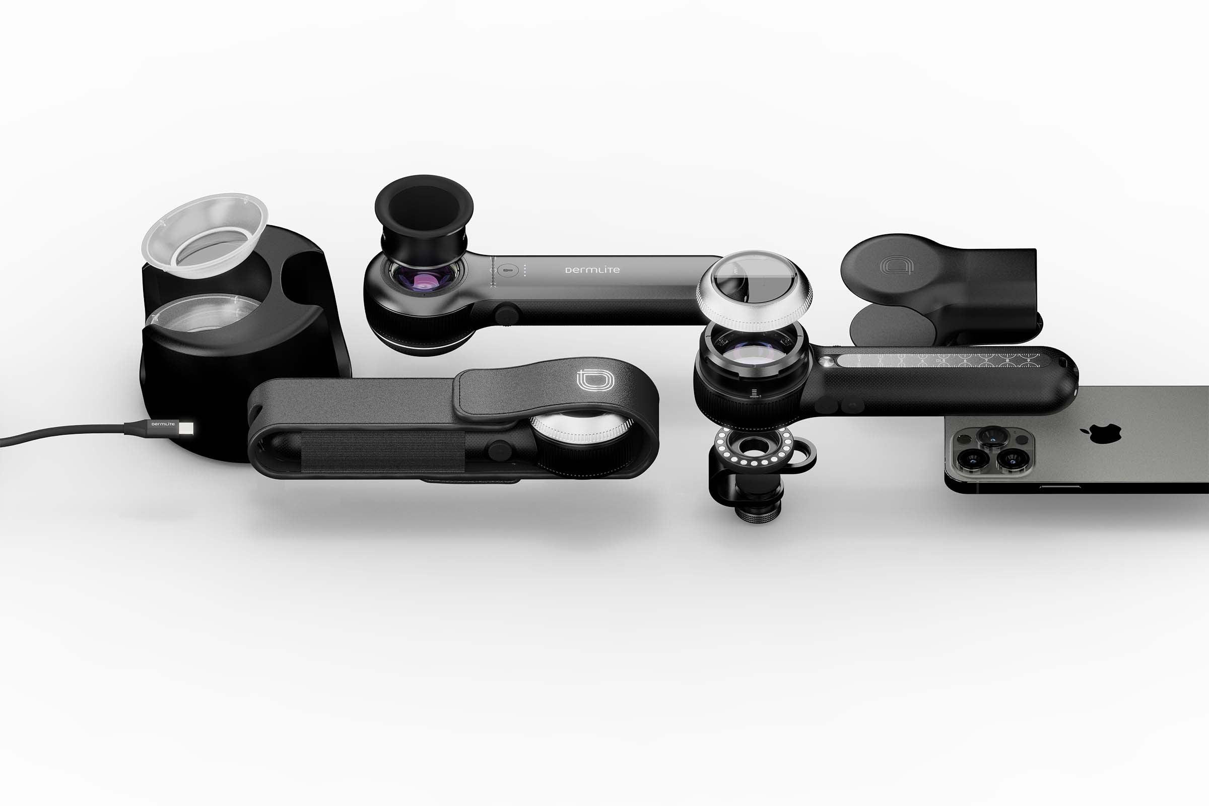
DL5 includes:
- handheld dermatoscope with 10x magnification, variable polarization, white/PigmentBoost/UV LEDs for dermoscopy, torch LED
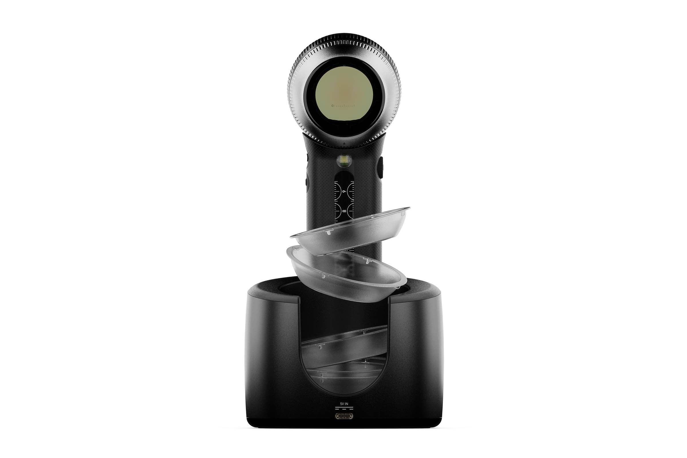
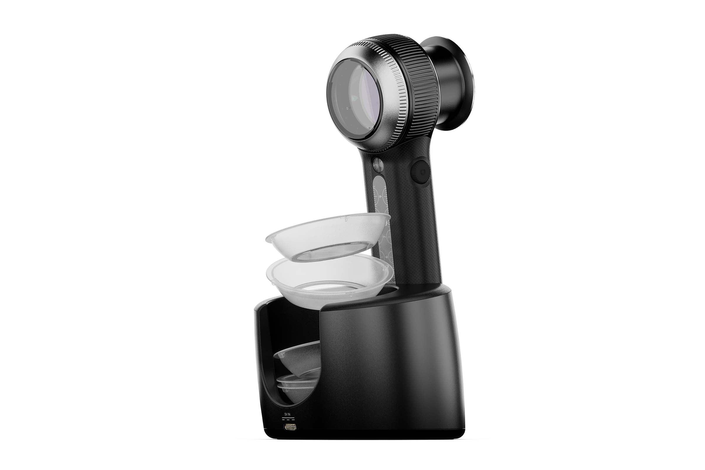
- desktop charging base with IceCap storage
- 2 m USB-C to USB-C cable
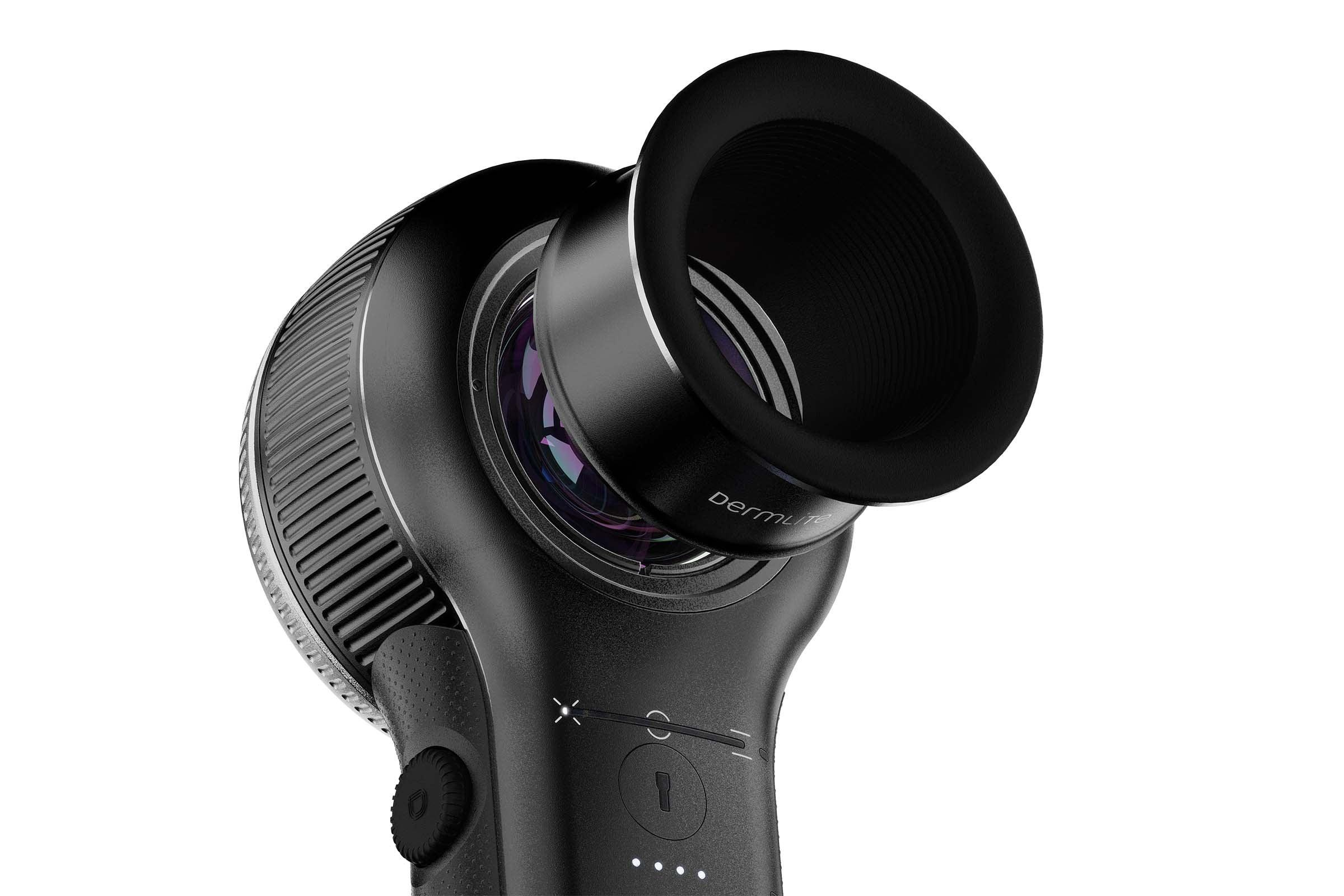
- eyepiece
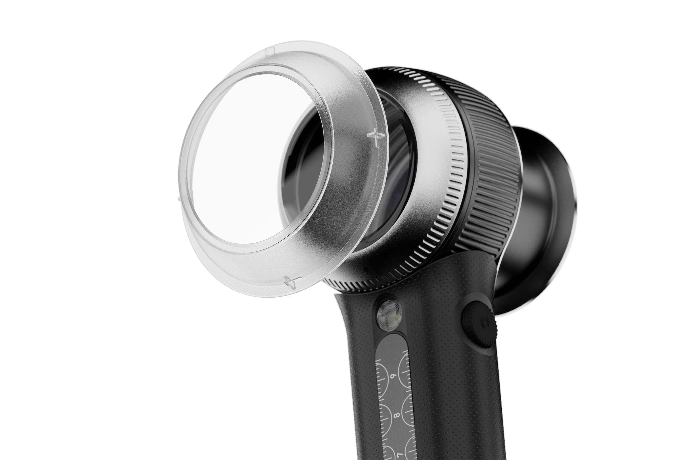
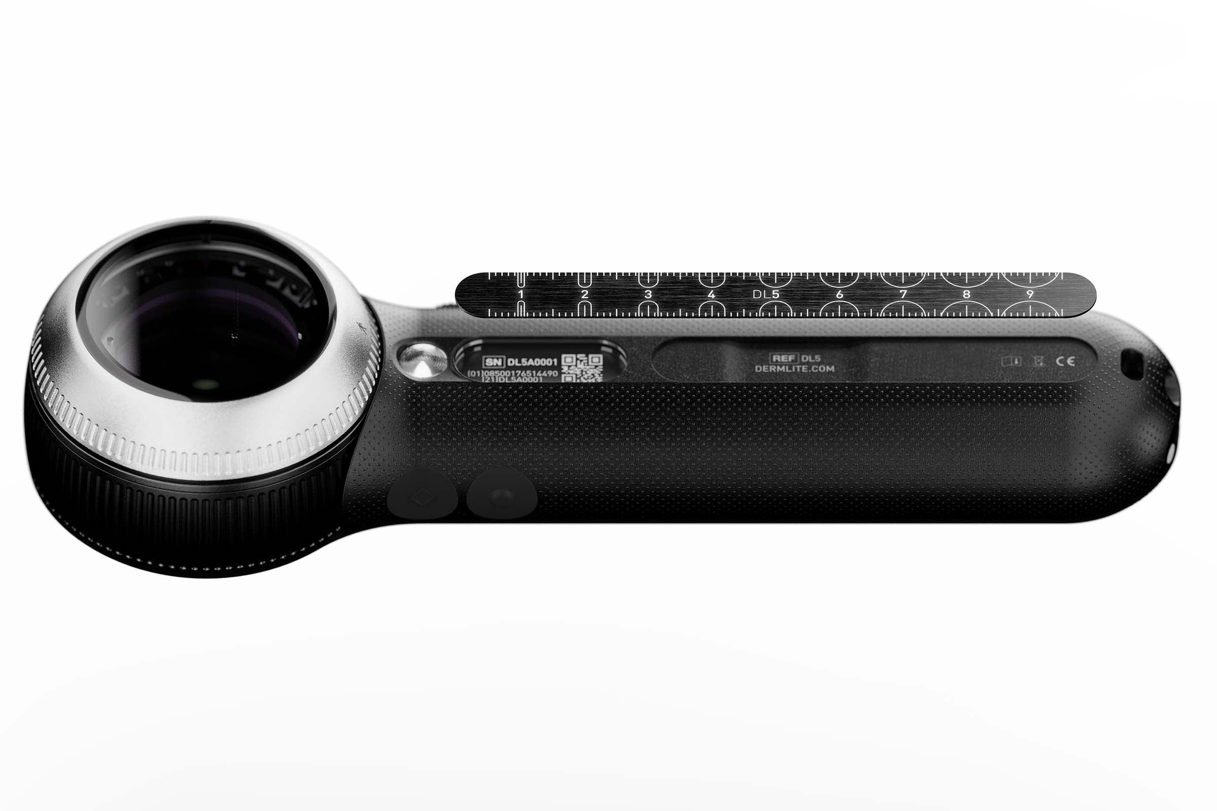
- magnetically attached stainless steel ruler
- leather belt pouch
- plastic belt holster
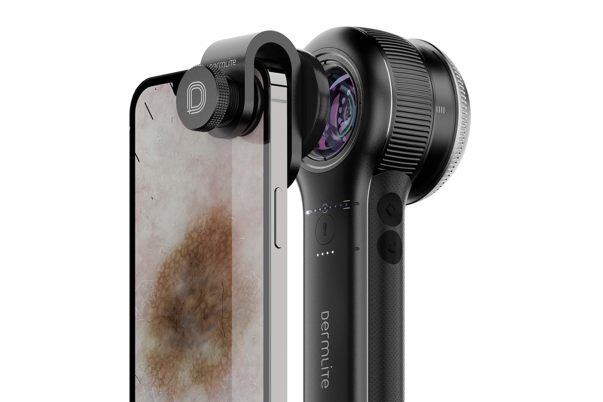
- universal smartphone connector MCC
- IceCap® 5-piece sample supply
- microfiber cleaning cloth
Size & mass: 182 x 58 x 36 mm, 285 g
Instructions for Use (PDF)
Compare to other pocket dermatoscopes
Note: Prior to using this product, please read the battery manufacturer'sMaterial Safety Data Sheet
Couldn't load pickup availability
More than a device.
Your daily companion that sees deeper.
DermLite is the world’s most popular dermatoscope for a reason. It's because we create our products because we’re curious, we listen, we think differently, and we want to inspire the world of dermoscopy. And to help make skin a little healthier, more beautiful. If you're on board, DL5 is your reliable companion. Your personal superpower.
DL5 is the result of 25 years of asking questions, thinking differently, and caring about the tiniest detail. It reflects global collaboration with visionaries in skin diagnostics and all those who love dermoscopy — or those who want to learn to love it.
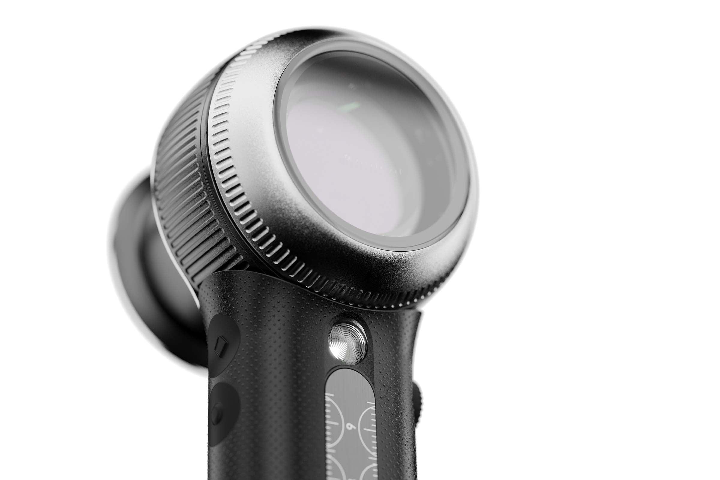
DL5 is your magic eye that shows you what others miss — crystal clear. One twist, and DL5 takes you deep into the skin. Across nine levels of polarization, brightness, and color. Into the fascinating world of UV dermoscopy.
Get inspired — by true 10x magnification, a finely tuned 32 mm optical system, and a design so well thought out you’ll forget it’s even there. Let’s go.
DermLite dealers
Delivery time
Due to a high volume of orders, the current delivery time is 3 weeks

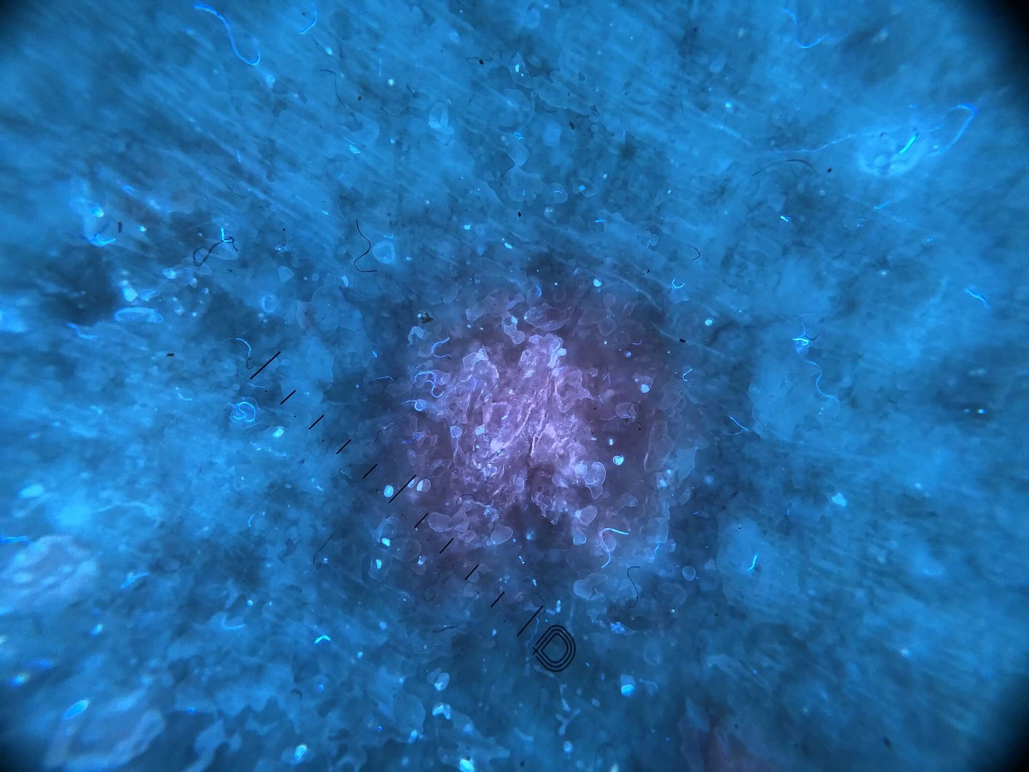
A new dimension of dermoscopy
DL5 brings UV fluorescence exactly where it matters most: into everyday diagnostics.
With 365 nm UV light at the press of a button — combined with true 10× magnification — it reveals details others simply don’t see.
Subtle fluorescence. Porphyrins. Early changes. And suddenly, everything becomes clearer.
Higlights at a glance
Taking optics a level further
A 32 mm lens system with true 10x magnification reveals even the finest details – crystal clear, whether viewed with the naked eye or on a smartphone. Thanks to variable polarisation, you can explore the depths of the skin with seamless clarity. The DL5 is optimised for what matters most: confidence in what you see. Why not play a few levels above everyone else?
Simply more hygienic. Because it’s the right thing to do.
Switch instantly between contactless and contact-based examinations—with or without an autoclavable contact plate. Our unique IceCap® system protects your patients, your team, and you with disposable contact glass caps—quickly, hygienically, and safely. Because sometimes the right choice is the simple one.
Light that thinks like you
Nine levels of brightness. Nine colour temperatures. Polarised. Non-polarised. Parallel polarisation, if you only need the surface. UV fluorescence, if you want to take things further. DL5 adapts to your vision – and your thinking. From first impression to final decision.
Your superpower. Everyday.
The DL5 fits naturally in your hand. No edges, no gaps – just clarity. It feels good, looks good and makes your everyday life easier. Whether you’re looking through the viewfinder or documenting with your smartphone – everything is exactly where it should be. The elegant charging station keeps it charged and organised – including space for your IceCaps. And if anything does go wrong: a 5-year warranty and personal service from Bavaria are included.
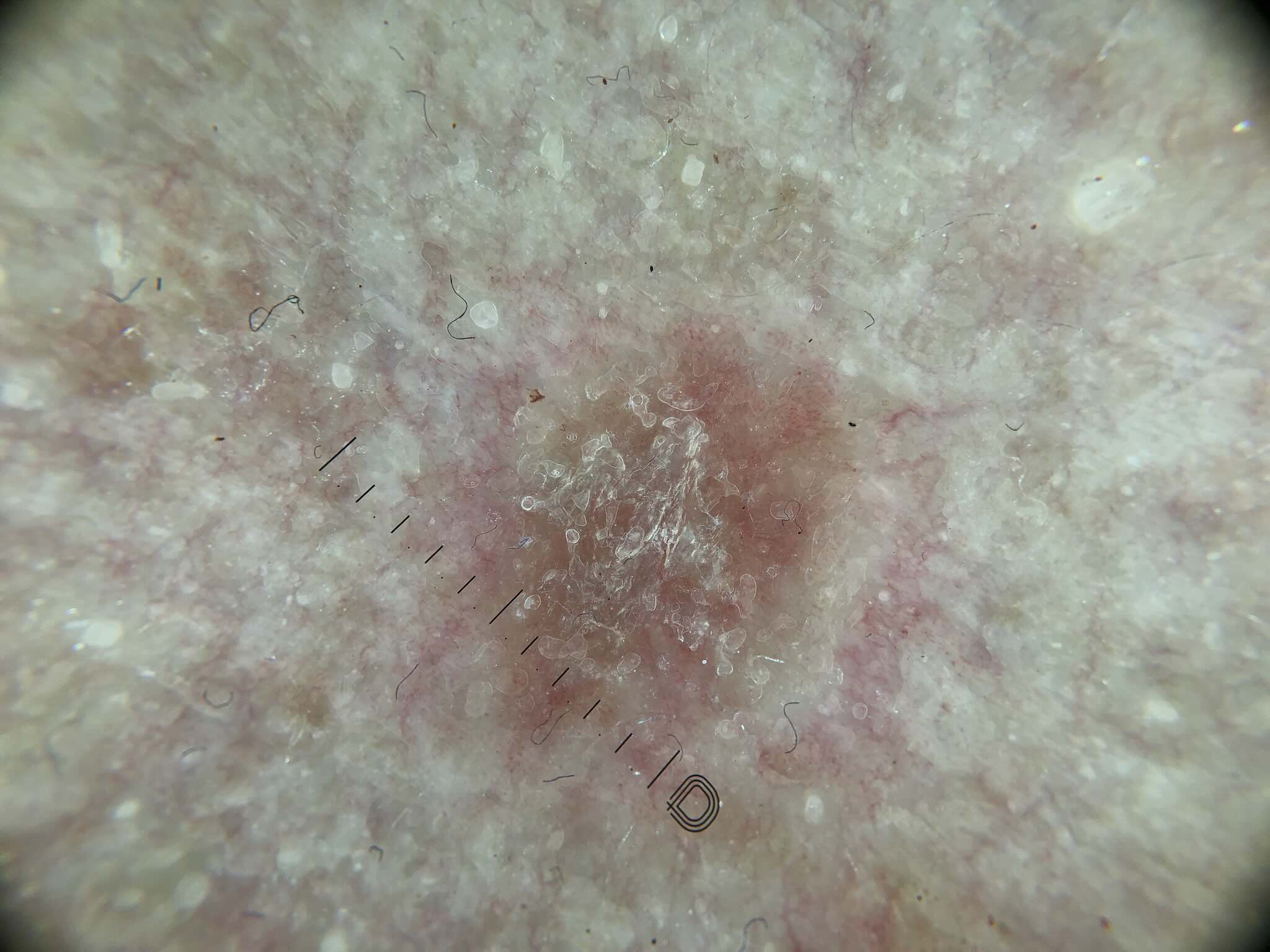
Psoriasis Polarized vs UV
| Feature | DermLite DL5 | Other Devices |
|---|---|---|
| Polarization | Full control across the polarization spectrum via scroll wheel | On/Off – no depth modulation |
| UV Mode | Integrated, 365 nm, clinically validated | Often not available |
| Clinical Illumination | Ultra-bright LEDs for a quick overview and hard-to-reach areas | Often not integrated |
| Magnification & Optics | 32 mm lens, true 10× – razor-sharp | Lower magnification, less detail |
| Design & Ergonomics | Balanced, grippy, made for your hand | Bulkier, more technical, less handy |
| Compatibility | Smartphone adapter included | Optional adapter required |
| Charging | Desktop charging station + USB-C – flexible | USB cable only |
| Included Accessories | Ruler, IceCaps, adapter, holster, more | Fewer or optional accessories |


Publications on UV dermatoscopy using the DL5
-
Systematic Review of UV Dermoscopy
Go to publicationApplications of Ultraviolet and Sub-ultraviolet Dermatoscopy in Neoplastic and Non-neoplastic Dermatoses
A Comprehensive Overview of the Applications of UV and Sub-UV Dermatoscopy in Benign and Malignant Skin Lesions.
-
Fordyce spots under UV dermoscopy
Go to publicationDifferentiating Fordyce Spots from Similar Lesions Using Ultraviolet-Induced Fluorescence Dermatoscopy
A Retrospective Study on the Differentiation of Fordyce Spots and Similar Lesions Using UVFD.
-
Yellow-green luminescent concretions in trichobacteriosis
Go to publicationUltraviolet Reflectance Dermatoscopy of Trichobacteriosis Axillaris
UVFD reveals characteristic peripilar luminescent concretions in trichobacteriosis axillaris
-
UV Therapy for Pityrosporum Folliculitis
Go to publicationUV Dermoscopy for the Diagnosis of Pityrosporum Folliculitis
UVFD as a diagnostic tool to aid in the rapid diagnosis of this common but often overlooked condition. -
Scabies under UV light
Go to publicationScabies Mites Glow Bright Green Under UV Dermatoscopy
Scabies mites fluoresce green under 365 nm light—a new visual feature for diagnosis. -
Yellow-green luminescent concretions in trichobacteriosis
Go to publicationUltraviolet Reflectance Dermatoscopy of Trichobacteriosis Axillaris
UVFD reveals characteristic peripilar luminescent concretions in trichobacteriosis axillaris.